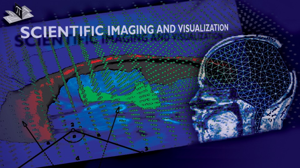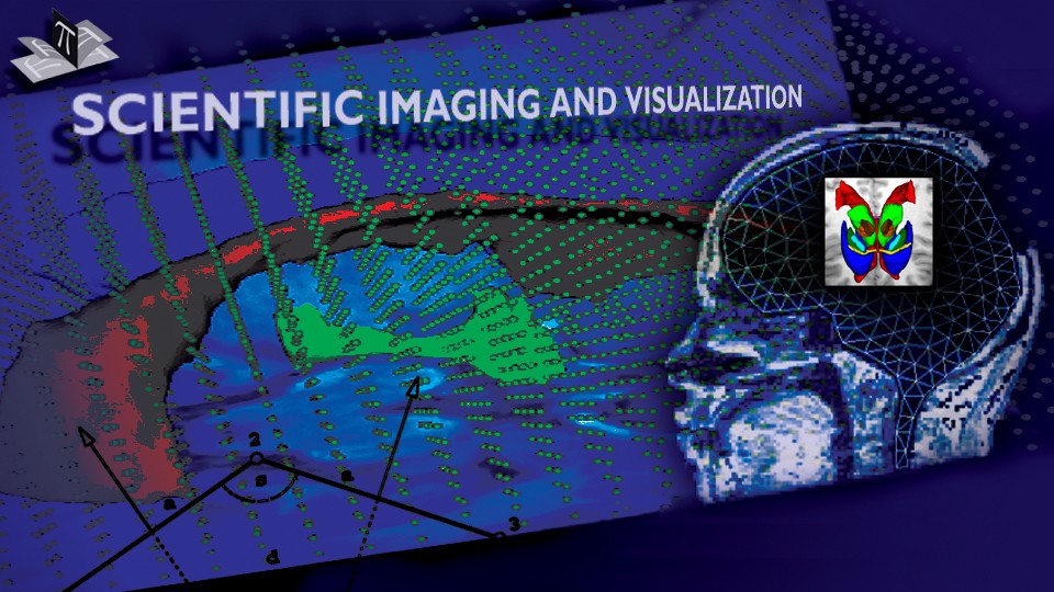


Inverse 3D FEM Analysis of a Brain Surgery Simulation

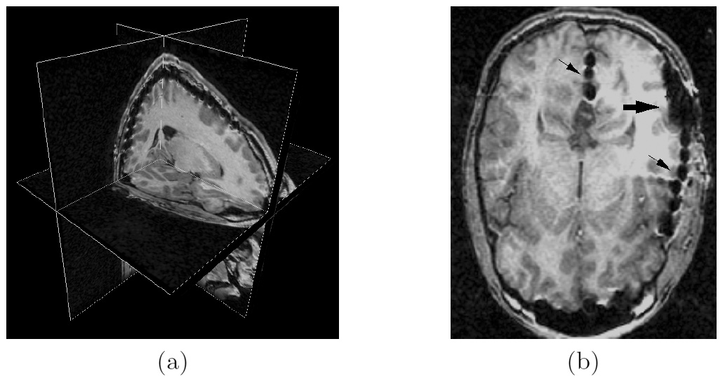
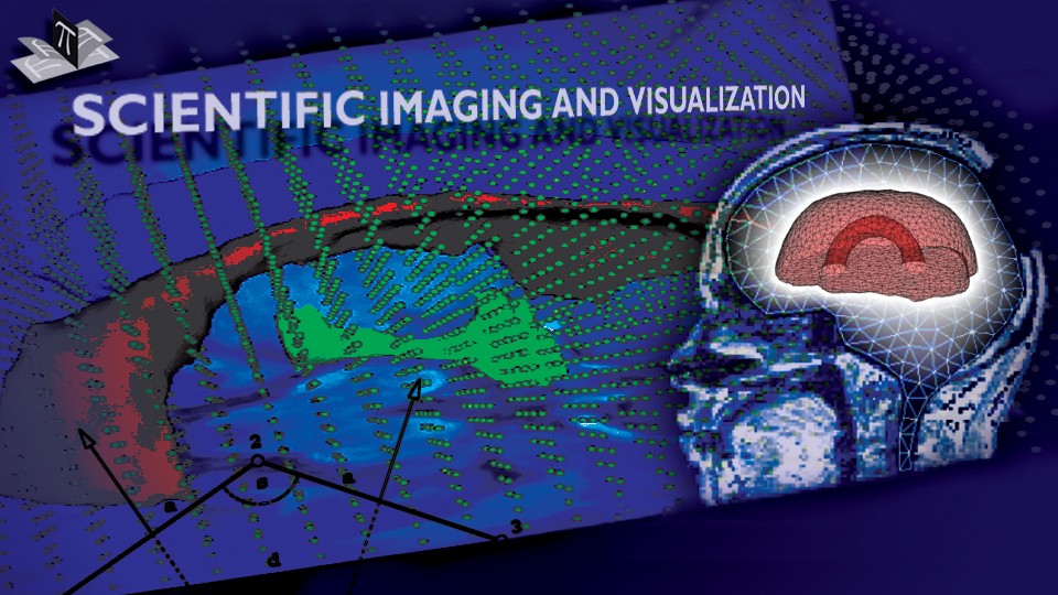
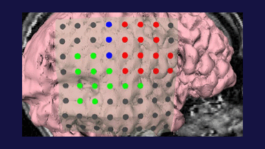
Image Guided Constitutive Modeling (IGCM) is a novel approach to development of reliable constitutive models of the mechanical behavior of the in-vivo human brain tissue. We propose to take the MR or CT scan of a brain response to ventriculostomy. Image-derived displacement fields are then used by 3D inverse analysis to develop the constitutive models of the brain tissue. In this project, the IGCM is demonstrated on the silicone brain phantoms (Fig. 1) closely simulating the in-vivo brain geometry, mechanical properties and boundary conditions.
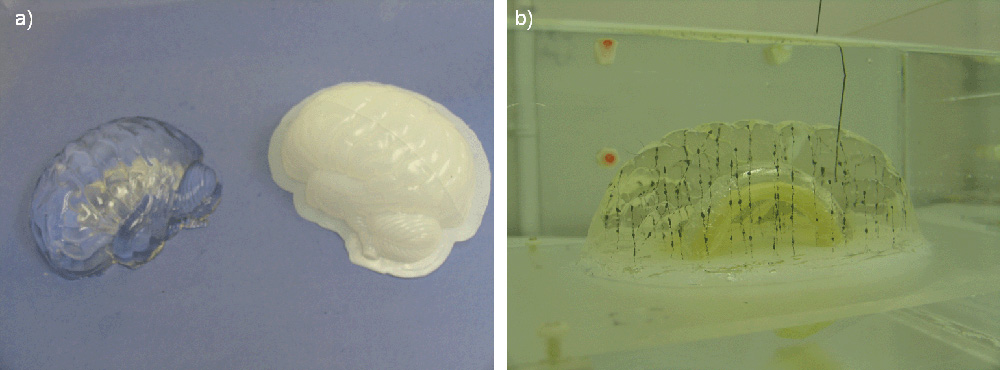
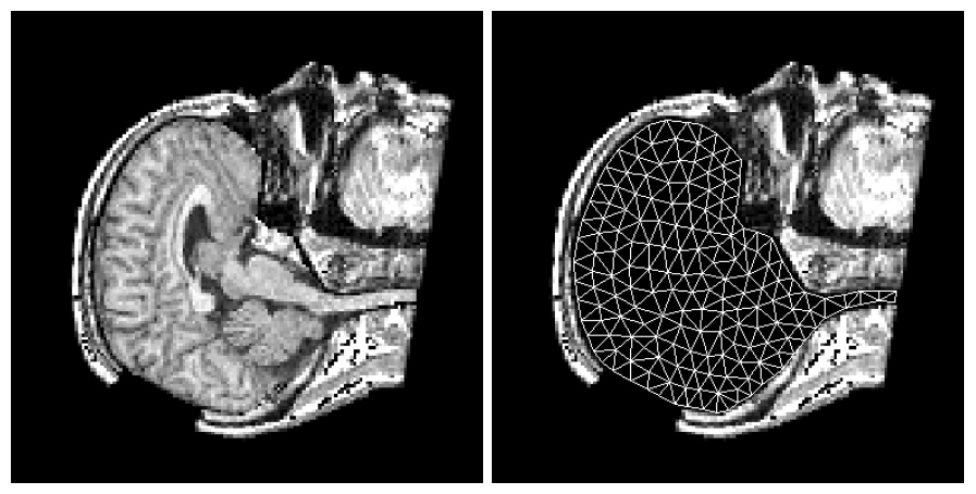
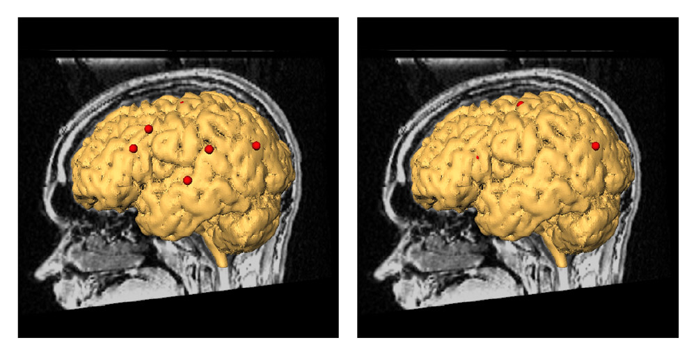
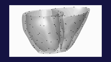
Figure 1: (a) Brain phantom (left) and plastic mold (right). (b) Brain phantom with validation beads. The figure is from [1] and it is used with permission; Copyright © 2006 Springer Berlin / Heidelberg; All rights reserved.
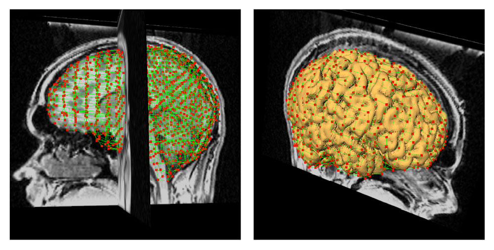
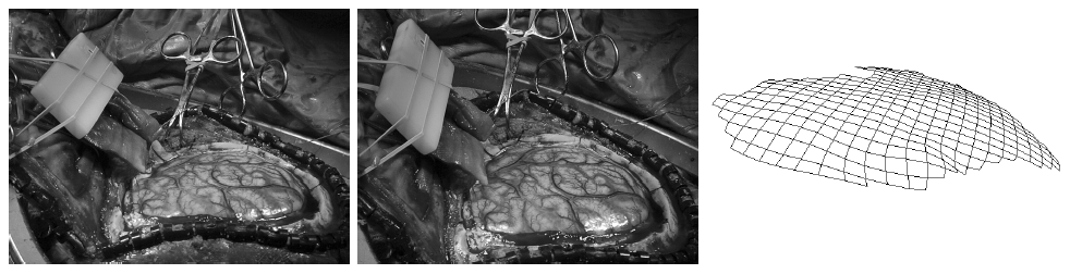
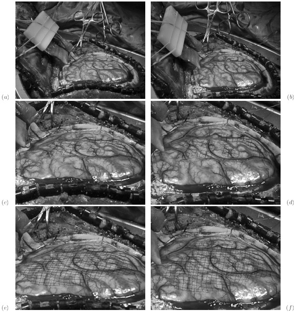
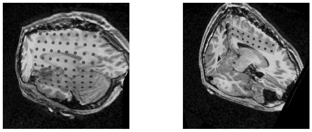
The ventriculostomy was simulated by consequently inflating and deflating the internal rubber membrane. The phantom was imaged in a CT scanner before and after deformation. Fig. 1 shows the experimental setup while Fig. 2 shows the schematic kinematics of the experiment.
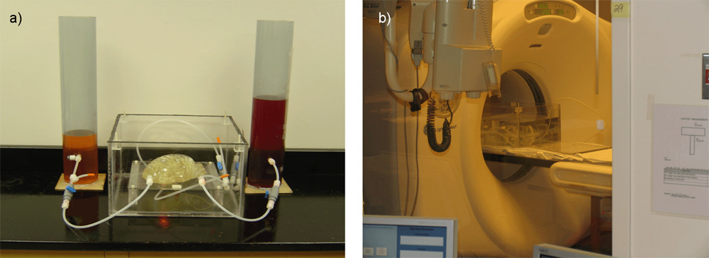
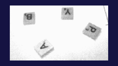
Figure 2: (a) Experimental setup. (b) Experimental setup in the CT scanner. The figure is from [1] and it is used with permission; Copyright © 2006 Springer Berlin / Heidelberg; All rights reserved.
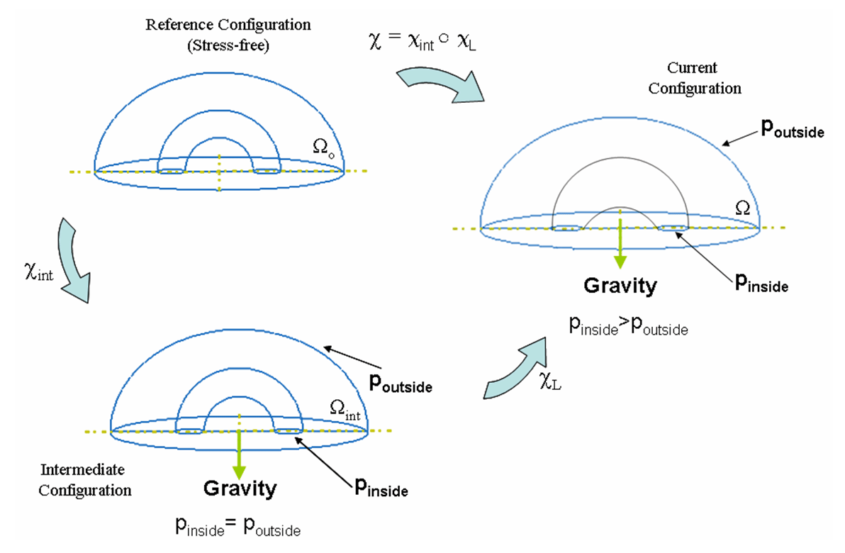
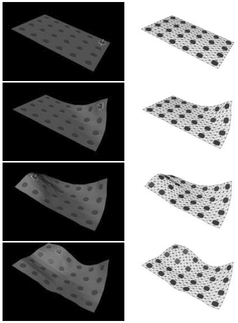
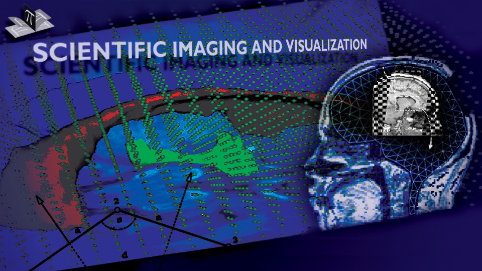
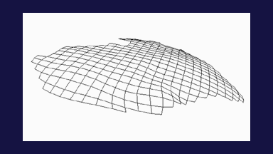
Figure 3: Schematic kinematics of the experiment. The figure is from [2] and it is used with permission; Copyright © 2006 Springer Berlin / Heidelberg; All rights reserved.
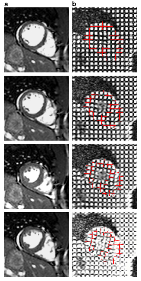
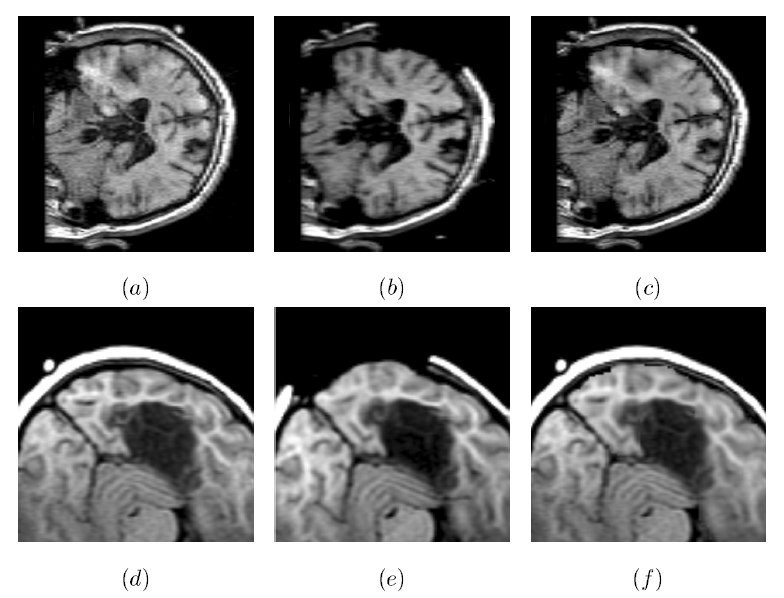
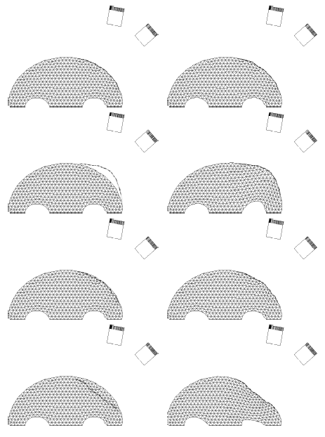
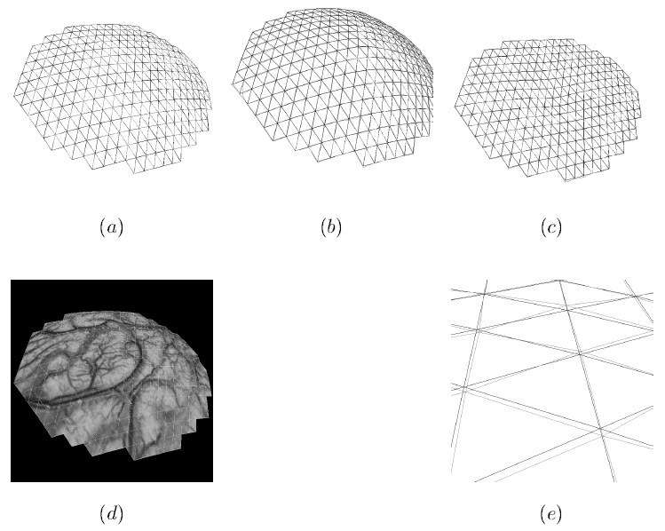
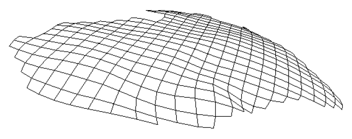
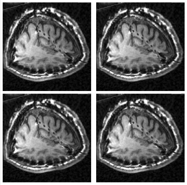
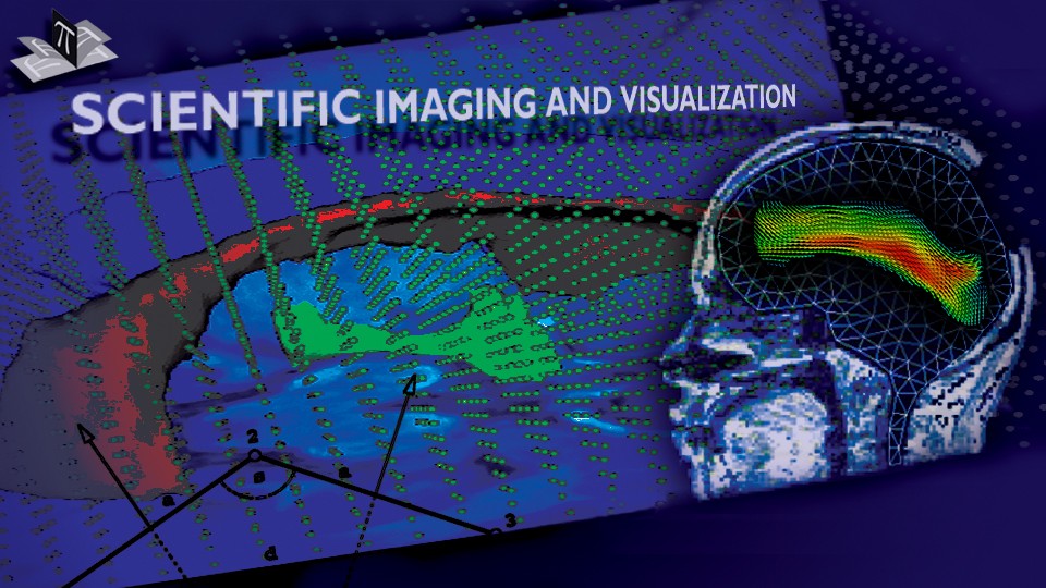
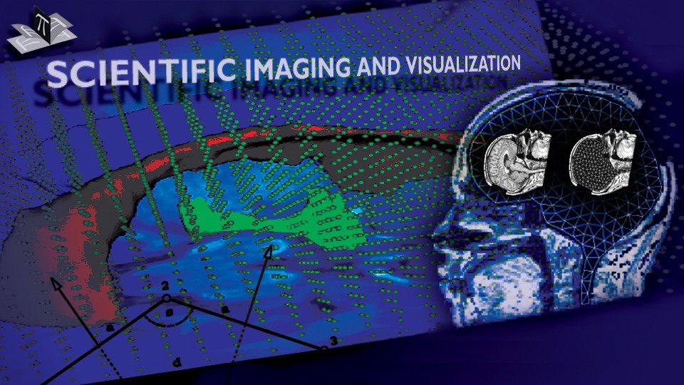
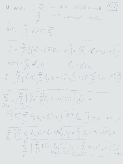
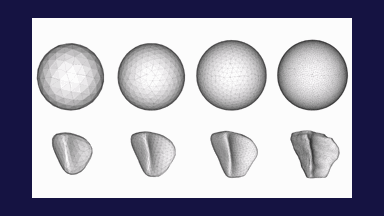
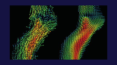
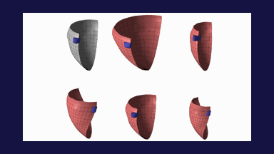
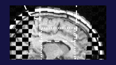
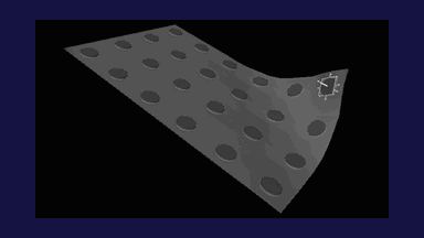
The obtained images were analyzed to derive displacement fields, which was followed by Inverse Finite Element Analysis to obtain the parameters of the neo-Hookean and Ogden elastic models for the phantom material. Fig. 4 shows the phantom surface mesh before and after the deformation, while Fig. 5 shows a cross-section through the 3D CT scan and FEM model before and after the deformation. The calculated mechanical properties were consistent with those in the literature and those obtained from the independent uniaxial compression tests, providing preliminary justification for the future application of the IGCM to in-vivo brain tissue.
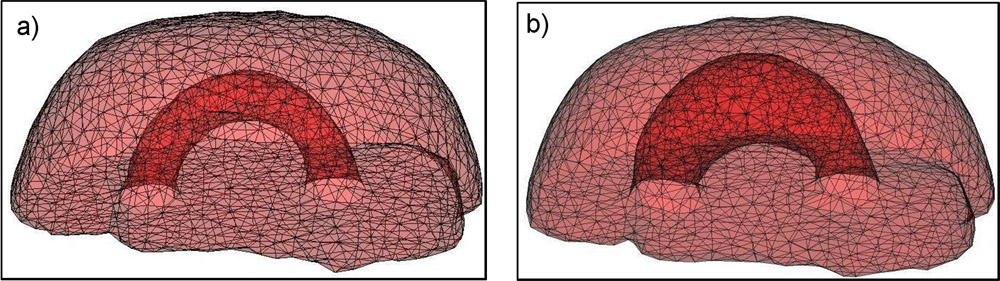
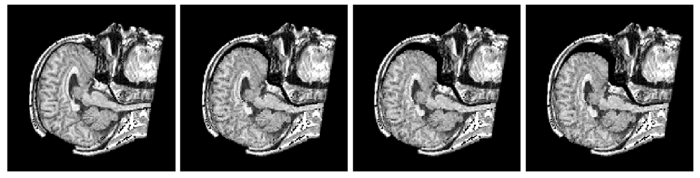
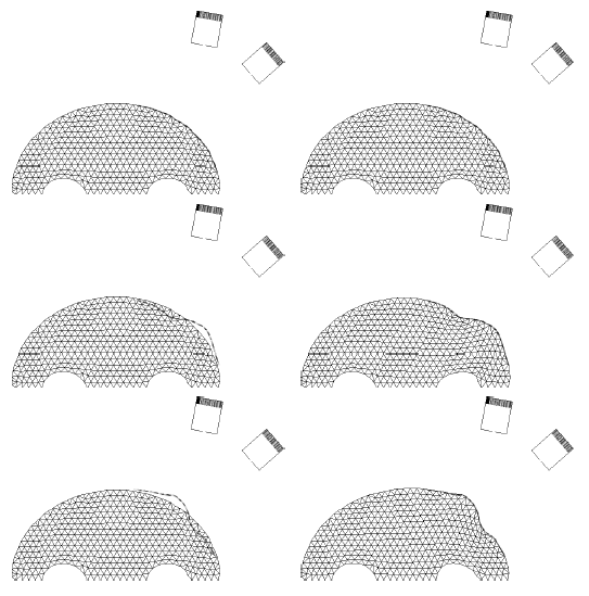
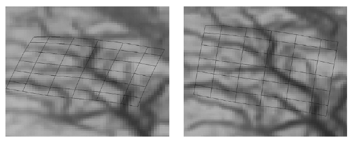
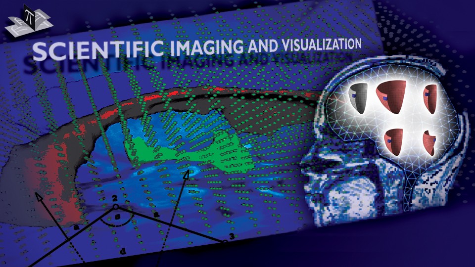
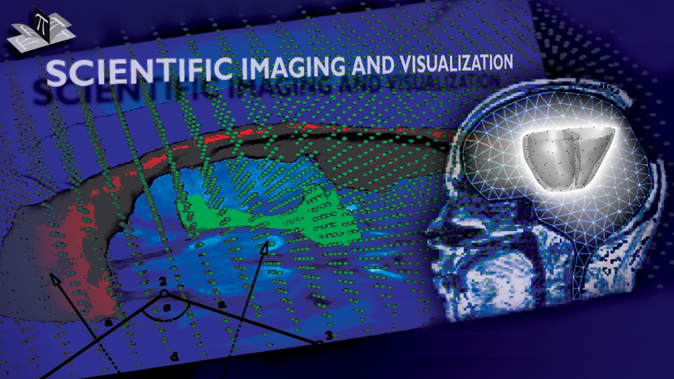
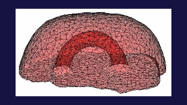
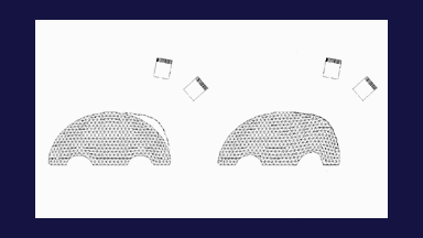
Figure 4: Surface mesh for (a) non-deformed and (b) deformed phantom. The figure is from [1] and it is used with permission; Copyright © 2006 Springer Berlin / Heidelberg; All rights reserved.
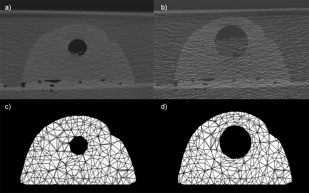
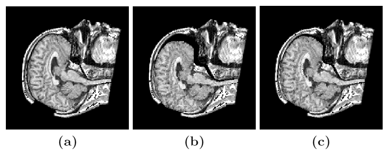
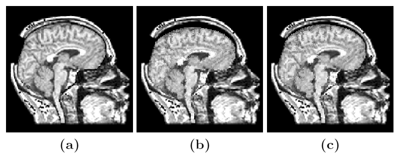
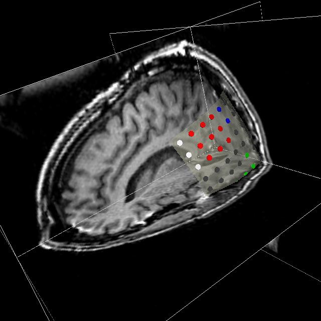
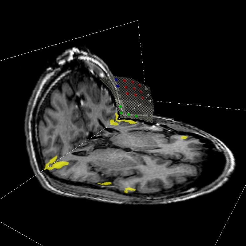
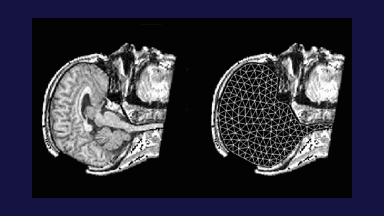
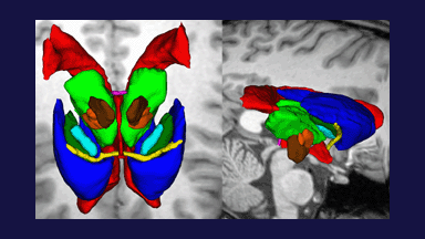
Figure 5: A scanned image slice (a) before and (b) after the deformation. The corresponding slices of the finite element meshes are shown in c) and d), respectively. The figure is from [1] and it is used with permission; Copyright © 2006 Springer Berlin / Heidelberg; All rights reserved.
References:
[1] Puzrin, A., Ozan, C., Germanovich, L., Mukundan, S., Skrinjar, O., "Inverse 3D FE Analysis of a Brain Surgery Simulation", Computational Biomechanics for Medicine, Copenhagen, Denmark, pp. 75-84, October 2006.